Deep Vein Thrombosis – Essential Guide & Resources
When dealing with deep vein thrombosis, a condition in which a blood clot forms in a deep vein, most often in the leg. Also known as DVT, it can quickly become dangerous if the clot breaks free and travels to the lungs. Understanding this condition early can save lives and prevent long‑term damage.
One of the core players in DVT is the blood clot, a semi‑solid mass of fibrin, platelets, and red cells that blocks blood flow. The clot’s size and location dictate how severe the symptoms become. Deep vein thrombosis frequently shows up as swelling, pain, or a warm feeling in the affected limb, but many cases hide without obvious signs. Knowing the symptoms, such as leg discoloration, tenderness, or sudden shortness of breath if a clot travels, helps you catch it before complications arise.
Key Factors That Push DVT Into Action
The likelihood of developing DVT spikes when certain risk factors, like prolonged immobility, recent surgery, cancer, or inherited clotting disorders, are present. Long flights, bed rest after an operation, or even sitting at a desk for hours without moving can let blood pool and clot. Age, obesity, and smoking add extra pressure on the veins, making it easier for clots to form. By recognizing these triggers, you can take simple steps—like periodic leg stretches or compression stockings—to lower your risk.
When a clot has already formed, doctors turn to anticoagulant therapy, medications such as heparin, warfarin, or newer direct oral anticoagulants that thin the blood and prevent clot growth. The goal is two‑fold: stop the existing clot from expanding and stop new clots from appearing. Treatment duration varies; some patients need a few weeks, while others require months of therapy based on the underlying cause. Monitoring blood levels and adjusting doses are essential to avoid bleeding complications.
Beyond medication, lifestyle tweaks can reinforce medical treatment. Keeping active with low‑impact exercise, maintaining a healthy weight, staying hydrated, and quitting smoking all support vein health. For people at high risk, doctors may recommend mechanical measures like intermittent pneumatic compression devices or graduated compression stockings. These tools mechanically assist blood flow, reducing the chance a clot will form in the first place.
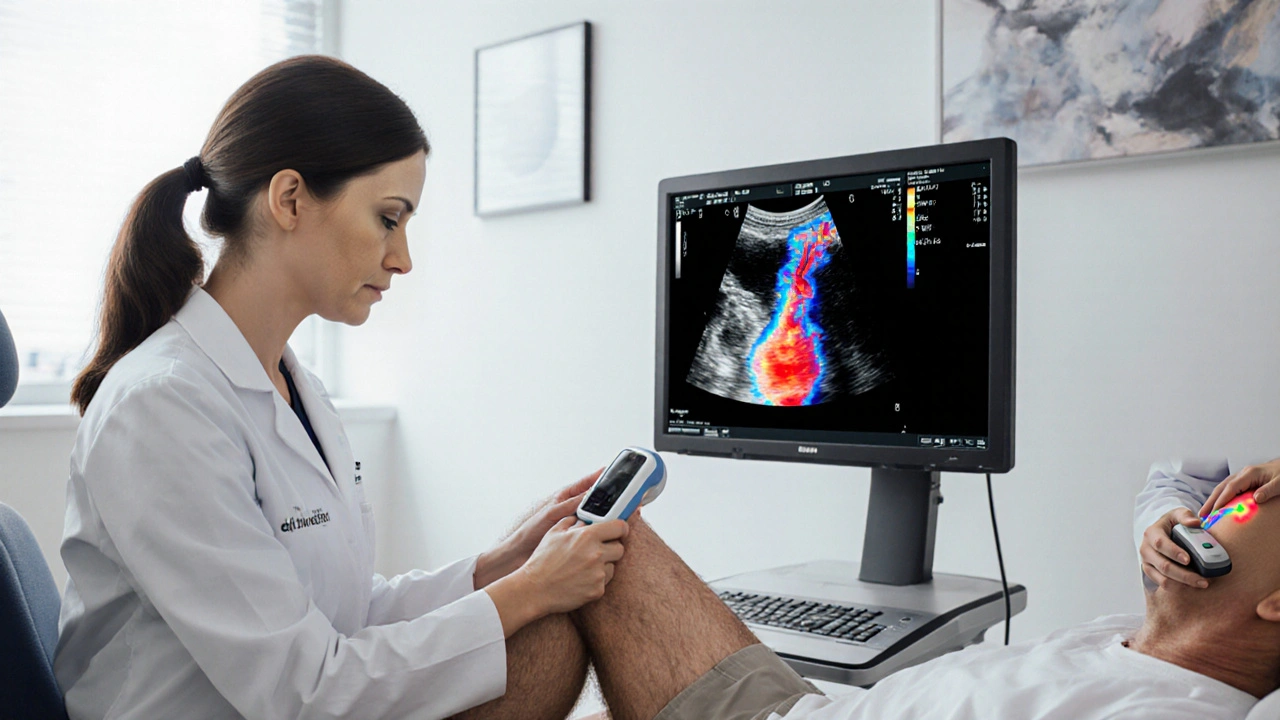
Diagnosis usually starts with a clinical exam, followed by imaging such as duplex ultrasound, which visualizes clot location and size. In ambiguous cases, D‑dimer blood tests help rule out clotting activity. Early and accurate diagnosis lets clinicians choose the right anticoagulant regimen and decide whether more aggressive interventions, like catheter‑directed thrombolysis, are needed.
The articles below dive into each of these topics in detail—ranging from how to spot hidden DVT symptoms, to choosing the right anticoagulant, to practical tips for prevention during travel or recovery from surgery. Browse the list to get actionable insights that match your situation and stay ahead of this silent but serious condition.