Venous ultrasound is a non‑invasive imaging technique that uses high‑frequency sound waves to visualize veins, evaluate blood flow, and detect clots, characterized by real‑time compression testing and Doppler velocity assessment.
TL;DR
- Venous ultrasound uses compression and Doppler to spot clots in minutes.
- It is >95% sensitive for proximal DVT and has no radiation.
- CT venography or MR venography are reserved for equivocal cases or pelvic veins.
- Positive scans trigger immediate anticoagulation; negative scans can be ruled out with a low‑risk clinical score.
- Point‑of‑care ultrasound lets emergency physicians confirm DVT at bedside.
How the Scan Works
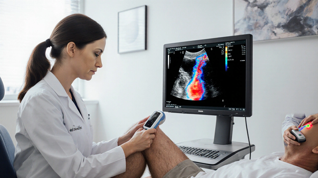
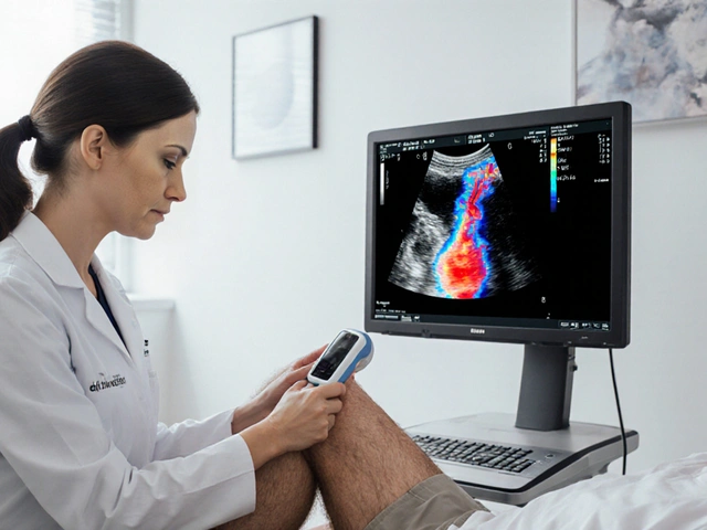
Two core principles drive the exam. Compression ultrasound is the act of pressing the transducer over a vein; a healthy vein collapses completely, whereas a thrombus blocks that collapse. The second principle is Doppler ultrasound, which measures the frequency shift of sound waves reflected off moving red blood cells. A sudden drop in velocity or a turbulent signal hints at a blockage.
The machine sends pulses at 5-10MHz, captures echoes, and translates them into a gray‑scale image. The operator moves the probe along the femoral, popliteal, and calf veins, applying gentle pressure every few centimeters. Modern scanners also overlay colour flow maps, making the distinction between flowing and static blood instantly visible.
From Symptoms to Scan: The Clinical Workflow
Before the ultrasound, clinicians assess pre‑test probability using the Wells score, a point‑based system that weighs factors such as recent immobilisation, cancer, and calf swelling. A score < 2 points is considered low risk; in that scenario, a negative ultrasound can safely rule out DVT without further testing. Scores ≥2 points are moderate‑to‑high risk, prompting immediate imaging.
Once the decision to image is made, a bedside or radiology‑department technician follows a standardized protocol: start at the common femoral vein, compress each segment, sweep to the popliteal vein, then evaluate calf veins if the proximal study is negative but clinical suspicion remains high.
Imaging Protocols: Limited vs. Extended Exams
A "limited" study focuses on the proximal veins (femoral and popliteal) because >95% of clinically significant DVTs originate there. An "extended" exam adds the entire calf, which increases sensitivity for distal clot detection but adds 5-10minutes to exam time.
Emerging point‑of‑care ultrasound (POCUS) programs let emergency physicians perform a rapid limited scan themselves. Studies from 2023‑2024 show POCUS sensitivity of 93% and specificity of 96% when operators complete a focused training curriculum.
How Venous Ultrasound Stacks Up Against Other Modalities
| Modality | Sensitivity (proximal) | Specificity (proximal) | Radiation | Typical Cost (USD) |
|---|---|---|---|---|
| Venous ultrasound | 97% | 96% | None | 200-400 |
| CT venography | 94% | 92% | Ionising (≈8mSv) | 600-900 |
| MR venography | 96% | 95% | None (magnetic) | 900-1,200 |
The table shows why venous ultrasound remains first‑line: it matches or exceeds the accuracy of CT and MR, avoids radiation, and costs less. CT is useful when a patient cannot tolerate compression (e.g., severe pain) or when pelvic veins need evaluation. MR is reserved for patients with iodine contrast allergy or chronic kidney disease.
Reading the Scan: Normal vs. Abnormal Findings
A normal result demonstrates complete compressibility at every level, smooth lumen walls, and pulsatile arterial flow adjacent to the vein. Colour Doppler should show uniform laminar flow without turbulence.
Abnormal signs include:
- Partial or absent compressibility - the classic “soft tissue mass” sign.
- Intraluminal echogenic material that does not move with compression.
- Altered Doppler waveform: loss of phasicity with respiration, or a high‑resistance pattern.
- Collateral veins or vein enlargement upstream of the clot.
False‑positives can arise from muscle artefacts, external compression from a cast, or venous valves that appear as non‑compressible points. Skilled technologists differentiate these by scanning in both transverse and longitudinal planes.
Impact on Patient Management
When a scan confirms DVT, guidelines from the American College of Chest Physicians (2024 update) recommend immediate initiation of anticoagulation therapy, typically a direct oral anticoagulant for a minimum of three months.
Negative ultrasound in a low‑risk patient (Wells ≤1) allows safe discharge without further testing. High‑risk patients with a negative scan may undergo repeat imaging in 5-7days or proceed to CT venography if clinical suspicion persists.
Beyond treatment, ultrasound can be used to monitor clot resolution. Serial scans every 2-4 weeks show decreasing size, re‑establishment of compressibility, and restoration of normal Doppler flow, guiding decisions about duration of anticoagulation.
Related Concepts and Next Steps
Understanding DVT risk factors-such as prolonged travel, recent surgery, active malignancy, and inherited thrombophilia-helps clinicians decide when to order an ultrasound. The Wells score itself is a decision‑support tool that integrates these risk factors into a single number.
For patients with suspected pelvic or upper‑extremity thrombosis, the same ultrasound principles apply, but the probe must be positioned differently, and sometimes CT venography offers better anatomical detail.
Future advancements include AI‑driven image analysis, which can automatically flag non‑compressible segments and quantify thrombus volume. Early studies in 2025 suggest AI‑assisted scans reduce interpretation time by 30% and improve inter‑observer agreement.
Why venous ultrasound Remains the Gold Standard
Because it blends high accuracy, bedside availability, lack of radiation, and relatively low cost, venous ultrasound is the go‑to test for most clinicians. Its ability to provide immediate answers in the emergency department shortens hospital stays and accelerates life‑saving anticoagulation.
Frequently Asked Questions
How soon after symptom onset can an ultrasound detect DVT?
Most clots become visible within 24-48hours. Early scans (<12hours) may miss a fresh thrombus, so a repeat scan is advised if clinical suspicion stays high.
Is compression ultrasound painful?
The pressure is usually well tolerated; the probe is pressed just enough to flatten the vein. Patients with severe leg pain may need a gentle technique or an alternative imaging test.
Can ultrasound miss clots in the calf?
Distal (calf) clots are smaller and harder to compress, so sensitivity drops to about 80% in a limited study. An extended scan or a repeat exam after a few days improves detection.
When should CT venography be considered?
CT venography is useful when ultrasound is technically limited (e.g., large dressings, severe obesity) or when physicians need to visualise pelvic veins that are beyond the reach of a linear probe.
What role does AI play in modern DVT imaging?
AI algorithms can automatically assess vein compressibility, colour‑Doppler flow and even estimate clot volume. Early adopters report faster reads and fewer missed clots, but AI should augment-not replace-human expertise.


Post A Comment