Doppler Ultrasound: What It Is and Why It Matters
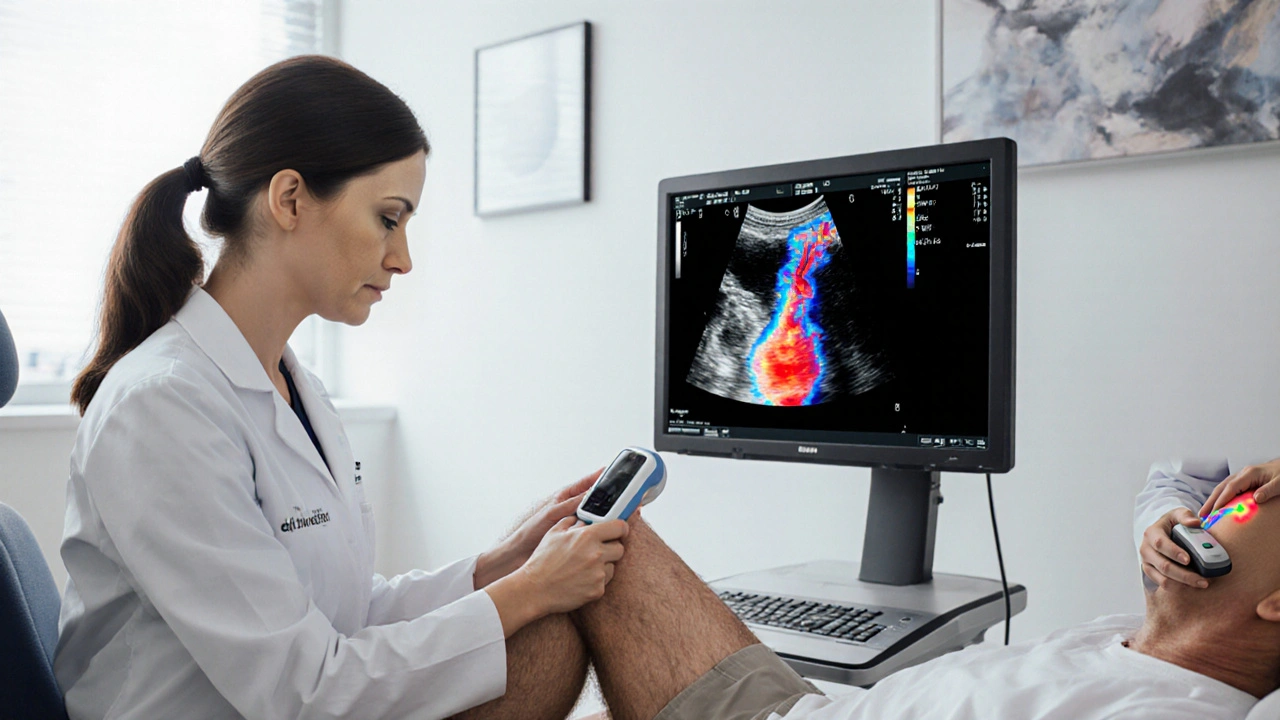
When working with Doppler ultrasound, a non‑invasive imaging method that uses sound waves to capture the speed and direction of blood flow in real time. Also known as Doppler sonography, it lets doctors see how blood moves through arteries, veins and organs without a single cut.
The technique is especially powerful for diagnosing vascular disease, any condition that narrows, blocks, or weakens blood vessels. When a vessel is narrowed, the Doppler pattern changes – louder, higher‑pitched sounds signal faster flow. By comparing those patterns to normal values, clinicians can pinpoint blockages that cause hypertension, leg cramps, or organ ischemia. In practice, doctors often pair Doppler findings with medication choices, such as the blood‑pressure drugs discussed in our hypertension guides.
Key Applications of Doppler Ultrasound
Another major arena is fetal monitoring, the use of ultrasound to evaluate blood flow in the placenta and umbilical cord during pregnancy. A healthy placenta shows steady, low‑resistance flow; abnormal spikes can warn of growth restriction or pre‑eclampsia. Expectant mothers reading our pregnancy articles will see how Doppler data influences decisions about delivery timing and nutritional support.
Beyond pregnancy, the method excels at blood flow measurement, quantifying volume and velocity of circulation in specific vessels. Surgeons rely on those numbers before procedures like kidney stone removal, because renal blood flow impacts healing. Patients managing chronic kidney disease often hear about Doppler scans in the context of medication adjustments, such as the renin‑angiotensin blockers featured in our drug reviews.
In the broader picture, Doppler ultrasound is one of several imaging modalities, techniques like MRI, CT and conventional ultrasound used to visualize internal structures. Unlike CT, it has no radiation, making it a first‑line tool for monitoring conditions that evolve over time – for example, the progression of Parkinson’s‑related tremor where blood‑flow changes in the brain can be a subtle clue. This safety profile also explains why it appears in articles about long‑term medication safety.
Semantic relationships tie these concepts together: Doppler ultrasound encompasses blood flow measurement, requires a specialized transducer, and produces data that influences treatment of vascular disease. Vascular disease, in turn, shapes the patterns clinicians interpret, while fetal monitoring demonstrates how the same physics help protect a developing baby. Finally, imaging modality choices depend on the clinical question, and Doppler often wins when speed and safety matter most.
Below you’ll find a curated list of articles that dive deeper into the drugs, conditions, and lifestyle tips that intersect with Doppler ultrasound findings. Whether you’re tracking blood pressure, planning a pregnancy, or managing a chronic disease, the insights here will help you understand how this simple scan fits into your overall health strategy.