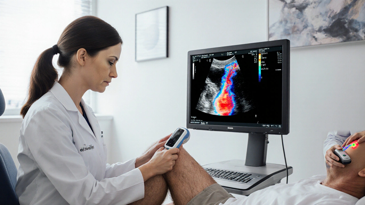
How Venous Ultrasound Diagnoses Deep Vein Thrombosis
Explore how venous ultrasound works, its role in diagnosing deep vein thrombosis, and how it compares to CT or MR venography for accurate, radiation‑free clot detection.
When talking about CT venography, a specialized computed tomography scan that visualizes veins using contrast material. Also known as CT venous angiography, it combines the speed of computed tomography, a cross‑sectional imaging technique that creates detailed 3‑D pictures of the body with a contrast agent, a dye injected into the bloodstream to highlight vascular structures. The result is a high‑resolution map of the venous system that helps clinicians spot clots, stenoses, or abnormal pathways.
CT venography is part of the broader field of vascular imaging, any imaging method that visualizes arteries, veins, and lymphatics. It often comes into play when doctors need to evaluate venous thrombosis, a blood clot forming in a vein that can cause swelling, pain, or life‑threatening complications. The procedure requires precise timing of the contrast injection, because the quality of the venous phase directly influences the ability to detect subtle clots. Imaging protocols also consider patient factors such as kidney function, since contrast agents can stress the kidneys.
Beyond clot detection, CT venography supports surgical planning, assesses traumatic injuries, and helps monitor disease progression in conditions like May‑Thurner syndrome. When you explore the articles below, you’ll see real‑world examples of how CT venography guides treatment decisions, what alternative imaging options exist, and tips for optimizing image quality. Whether you’re a patient curious about the test or a clinician looking for best practices, the collection ahead covers the practical side of CT venography and its related topics.

Explore how venous ultrasound works, its role in diagnosing deep vein thrombosis, and how it compares to CT or MR venography for accurate, radiation‑free clot detection.