Venous Ultrasound: What It Is and Why It Matters
When working with venous ultrasound, a non‑invasive imaging technique that uses high‑frequency sound waves to visualize veins. Also known as vascular sonography, it helps clinicians assess blood flow, detect clots, and guide treatments. Venous ultrasound covers both structural and functional aspects of the venous system, making it a first‑line tool for many vascular conditions.
Key Related Concepts
One of the most common reasons to order a venous ultrasound is to rule out deep vein thrombosis, a blood clot forming in the deep veins, usually of the legs. Early detection can prevent life‑threatening pulmonary embolism. The exam also relies on duplex ultrasound, a combined B‑mode and Doppler technique that shows vein anatomy and flow direction. Duplex provides the real‑time feedback needed to differentiate a harmless varicose vein from a dangerous thrombus. Together, these tools form the backbone of modern vascular imaging, the broader field of imaging methods that evaluate blood vessels.
Beyond clot detection, venous ultrasound is essential for diagnosing peripheral venous insufficiency, evaluating valve competence, and planning compression therapy. In patients with chronic swelling, the scan reveals reflux patterns and helps determine the severity of valve failure. This information guides decisions about graduated compression stockings, minimally invasive ablation, or surgical vein stripping. Because the test is painless, repeatable, and free of radiation, doctors can monitor disease progression over months or years.
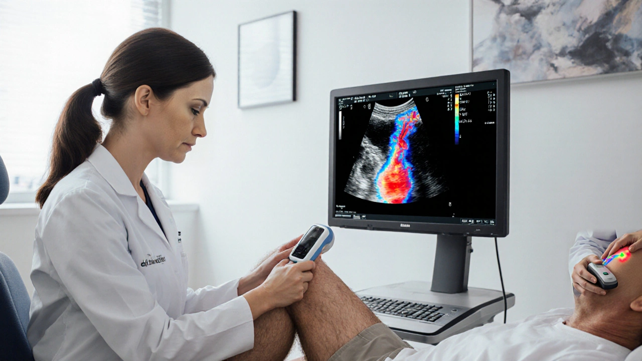
Technically, the procedure follows a clear workflow: a technician selects the appropriate transducer frequency, maps the superficial and deep venous segments, and applies color Doppler to assess flow. The resulting images are then interpreted against established criteria—such as the compressibility test for DVT or the augmentation test for reflux. Accuracy hinges on operator skill, probe positioning, and patient preparation. Modern machines now include AI‑assisted edge detection, which can highlight suspected thrombi in seconds, but a trained eye is still the gold standard.
Understanding how these pieces fit together prepares you for the range of topics covered in the articles below. Whether you’re curious about the latest DVT screening protocols, want a step‑by‑step guide to performing duplex scans, or need tips on interpreting vascular imaging reports, the collection offers practical, up‑to‑date information. Dive in to see how venous ultrasound shapes diagnosis, treatment, and long‑term care for vein‑related health issues.