Compression Ultrasound: A Practical Overview
When working with Compression ultrasound, a non‑invasive imaging method that uses sound waves while a cuff gently squeezes the leg to visualize blood flow and vein walls. Also known as compressed duplex scan, it helps clinicians see how veins react to pressure. Related tools include Deep Vein Thrombosis, a blood clot that forms in the deep veins of the leg and can be life‑threatening if it travels to the lungs, Duplex ultrasound, a standard ultrasound that combines B‑mode imaging with Doppler flow analysis to assess vein structure and circulation, Compression therapy, the use of graded elastic garments or bandages to improve venous return and reduce swelling, and Venous ulcer, a chronic wound that forms near the ankle when vein pressure stays high for a long time. Together, these entities form a network: compression ultrasound evaluates vein function, which in turn guides compression therapy; compression therapy reduces the risk of venous ulcer; and identifying DVT early changes treatment plans.
Why It Matters for Everyday Care
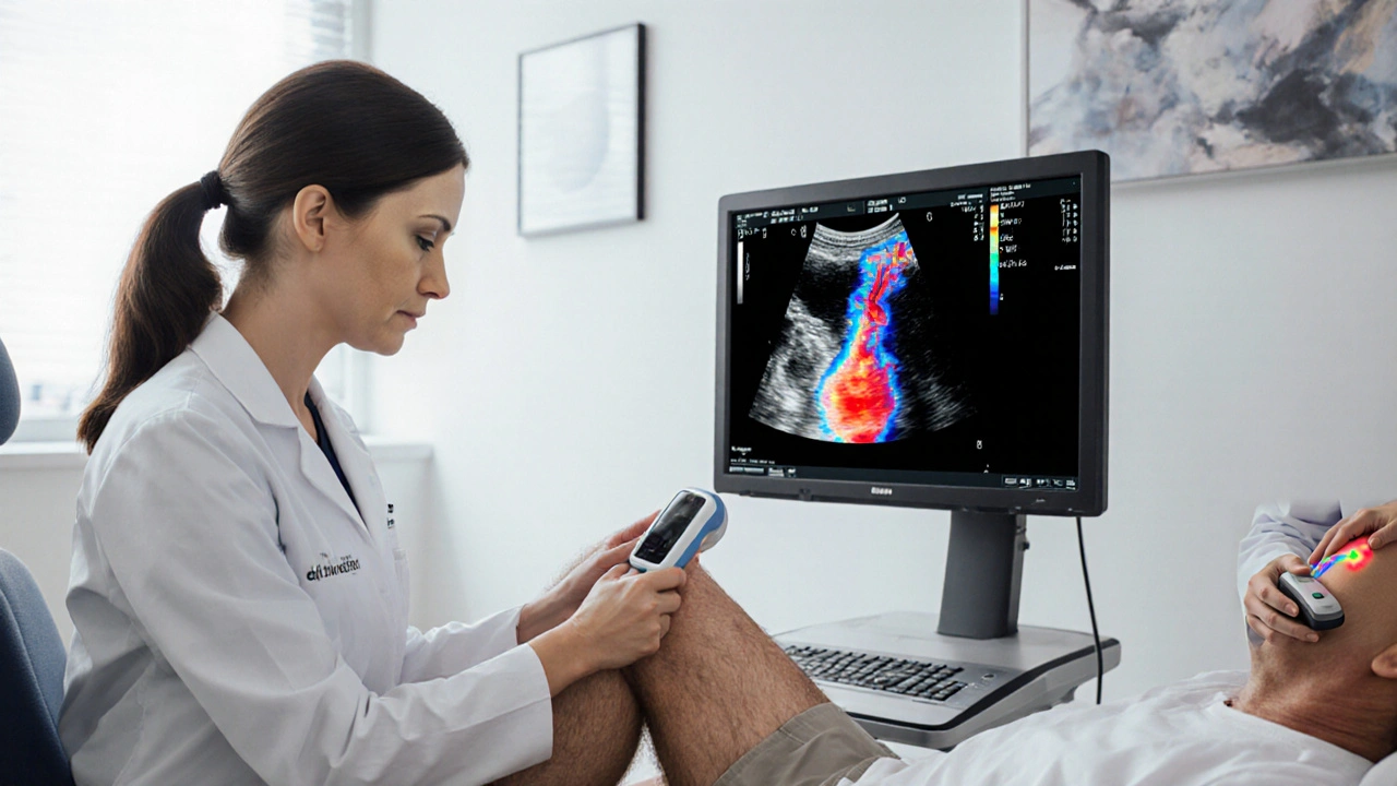
Think of compression ultrasound as the eyes that watch how a vein behaves under pressure. When a cuff squeezes the leg, the machine measures how quickly blood rushes back – that’s the “reflux” sign doctors look for. If reflux is present, the vein is likely weak, and a clinician may prescribe compression therapy to boost return flow. On the other hand, if the scan spots a sudden blockage, that points to deep vein thrombosis, prompting anticoagulant treatment instead of stockings. This dual role makes compression ultrasound a bridge between diagnosis and therapy. In our experience, patients with early‑stage venous ulcer benefit most when the test confirms that compression therapy will actually improve healing. The test also helps rule out arterial problems that could mimic vein issues, ensuring the right plan is applied. By pairing the imaging results with duplex ultrasound findings, doctors get a full picture of both structure and flow – a combo that beats guessing.
Below you’ll find a curated set of articles that dive deeper into each of these connections. From how compression ultrasound guides the choice of compression stockings, to real‑world cases of DVT detection, to tips for managing venous ulcers, the posts cover practical steps you can apply right away. Whether you’re a patient curious about why your doctor ordered the test, or a healthcare professional looking for clear explanations, the collection gives concrete insights that go beyond theory. Let’s explore the specifics together and see how this simple scan can shape better outcomes.